Patella luxation (Knee cap dislocation) is the seventh most commonly diagnosed canine orthopaedic condition in the UK. Its where the knee cap subluxes medially or laterally in dogs and sometimes cats. It does happen in humans too however the knee cap most commonly subluxes laterally (outside of the knee), whereas in animals its subluxes medially (inside of the knee).
The severity is dependent on the grade, which is categorised 1-4.
Luxation grades
| Grade 1 | Knee cap can be manually luxated but it returns to its normal position in the grove when released |
| Grade 2 | Knee cap luxates with stifle(knee) flexion or on manual manipulation and remains luxated with stifle extension or manual replacement occurs |
| Grade 3 | Knee cap luxated continuously. It can be manually replaced but re-luxates spontaneously when manual pressure is removed |
| Grade 4 | Knee cap luxated continuously and cannot be manually replaced |
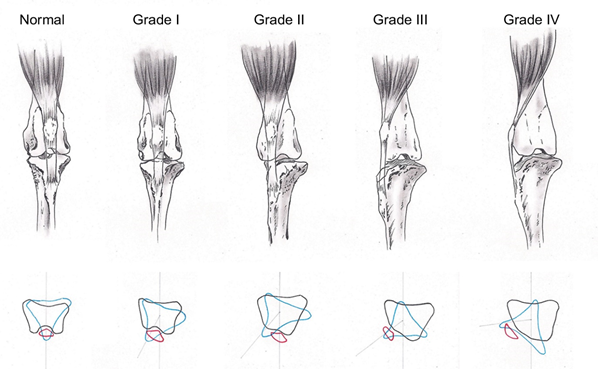
(Image from: Dona, Delle Valle and Fatone, 2018)
Developmental Causes:
- Patella luxation is not completely understood however it is thought the ‘Extensor Mechanism’ plays a key role in the development of the pathology. The quadriceps muscle group, the knee cap, the patella ligament and the bony prominence it attaches to (Tibial Tuberosity) are all part of the ‘Extensor mechanism’ of the stifle/knee. Any abnormality of this mechanism can lead, during the growth period, to bony changes of the femur (thigh) and tibia (shin) and subsequently cause patellar laxity.
- The thigh bone has a bony prominence for muscle attachment at the top, known as the femoral neck. The angle of this bony prominence can affect the ‘Extensor Mechanism’ because of the quadriceps muscle group passing and attaching onto it. The angle can affect the amount of torque the knee joint produces and can cause laxity in the mechanism.
- The knee cap sits in a grove known as the trochlear grove, which if shallow (trochlear hypoplasia) can cause patella luxation. The lack of depth in the grove is a developmental abnormality which is thought might be caused by the absence of physiological pressure on the cartilage of the grove itself.
- Malalignment of the femur and tibia can affect the ‘Extensor Mechanism’ due to changing the angle of the knee joint. This is known as ‘The anteversion angle’.
Patella luxation can be caused by trauma due to stretching of muscles and nerves however this is less common than developmental causes.
Which dogs are at risk of patella luxation?
- Small breeds are most susceptible, but research is showing large dogs are gradually being identified in veterinary practice.
- Dogs with a shallow groove where the knee cap sits, can make it more likely for the knee cap to move medially or laterally.
- Females are more commonly affected
- Neutered dogs have 3 times the risk of patella luxation
- Dutch Flat-coated Retrievers and Pomeranians
Symptoms:
- Intermittent or continuous lameness is displayed depending on the grade of luxation. This can range from a skipping pattern to bunny hopping, or putting no weight through the limb at all
- Hesitation or avoiding jumping off the sofa, the stairs or out of the car
- You might see the animal stretch the leg backwards to try and relocate the knee cap
- Their trot or walk may look quite stiff due to the affected ‘Extensor Mechanism’
Management:
Depending on the grade of the luxation will depend on the management. There is a risk of injuring the ‘CCL’ ligament with medial subluxing knee caps, therefore again management can be different. If just the knee cap is subluxing however, and no other structures are injured management Is usually standard across veterinary practices. A Grade 1 is usually managed through advice and physiotherapy, whereas grade 2,3,4 usually require surgical intervention. There are a few options for surgery however this is decided by your vet based on the cause of the patella luxation.
- Femoral Trochleoplasty – Deepening the shallow groove where the knee cap sits
- Tibial Tuberosity Transposition – The tibial tuberosity (the bony prominence where the quadricep tendon attaches) can be moved either towards the outside of inside of the knee joint
- Soft tissue release – this involves tightening and loosening certain muscles around the knee cap to reduce the subluxation
- Surgery can also combine all of these approaches or just two depending on the grading and the vets knowledge/experience
Physiotherapy after surgery:
- Control the inflammation – swelling is a natural response from the body following trauma, such as surgery. Its response in the early days is very normal because it provides a physiological response by bringing cells to the area to almost ‘clean’ it. But swelling which lasts more than 7-10 days can begin to make the joint stiff so cold therapy can help in preventing the swelling lasting longer than required.
- Activity restriction- This can also help control inflammation and usually involves crate rest for 8 weeks with the avoidance of slippery floors. Playing with other animals is not advised alongside, avoiding stairs and jumping from furniture. Your dog can be taken outside for the toilet but needs to be on a short leash.
- Restoring range of movement – The physiotherapist is trained to facilitate and therapeutically handle the limb to gradually get it moving through a comfortable range. This is done with the combination of pain relief and heat/cold therapy.
- Strengthening – this phase occurs when the pain has reduced. Exercises are prescribed tailored to the animal and include a wide range of programmes which can be done at home.
- Electrotherapy is also used to help with pain relief alongside reducing any muscle tightness.
Please get in contact with us if you think your dog has any of these symptoms and find out how we can help you.