TPLO and Physiotherapy for your dog

A Tibial Plateau Levelling Osteotomy (TPLO) is one of the most common surgeries I see in Physiotherapy clinic, so if your dog is due to have or has had a TPLO please do not panic! This blog is targeted at owners so you can learn about why a TPLO is needed and the role of Physiotherapy. I strongly feel owners need to feel educated and informed about their dog’s next steps so the right decisions can be made. So, let’s start from the beginning!

Why would my dog need a TPLO?

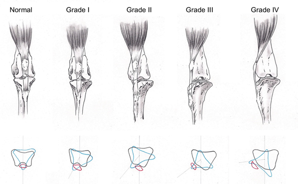
A TPLO is a type of surgery used to treat either a partial or fully ruptured Cranial Cruciate Ligament (CCL). When thinking about this in humans, a CCL is the same as an Anterior Cruciate Ligament (ACL) which works exactly the same. The CCL (or ACL) helps to stabilise the knee by preventing forward movement of the tibia on the femur (the shin bone and the thigh bone).

When you read about the cause of a CCL rupture you will see the term ‘degenerative changes’. Now I tend to dislike that phrase and something I refrain from using in my human practice, mainly because the wording can have negative or worrying connotations for patients. It sounds so much worse than what it means. I tend to use the analogy, ‘it’s like when your hair goes grey’ because it’s a ‘natural ageing process within the body’, so overtime the ligament can weaken. This process can also be accelerated by other factors, such as weight, breed, or conformation so in some cases dogs can be quite young and have this injury. Another added cause to a CCL injury is how the dog is anatomically made. In standing a dog will stand with roughly 110degrees of knee bend (on average), which means the CCL is always under strain. When there is strain or tension, intrinsic stress can occur in the joint and the risk of injury is higher, hence why younger dogs can sustain this injury.

What are the symptoms of a CCL rupture?

The symptoms overall are obvious to owners because your dog just won’t be their normal self. I have written the most obvious symptoms below.

  • Inability to put their full weight through the leg – pain and inflammation are common drivers to this because of the excessive movement of the tibia on the femur.
  • Stiffness on rising, getting out of bed, after walks or at the start and end of the day – this is usually due to the level of swelling within the joint but also the muscles around the joint becoming inactive and tight.  
  • Aggression to other dogs or sometimes people due to the level of pain.
  • Reluctance to go upstairs, jump in and out of the car or the sofa.

What is a TPLO?

A TPLO is one type of surgery used for this injury, it is widely performed and first described in 1993. It involves cutting into the bone called the tibial plateau (top of the tibia) and rotating it, meaning the tibia cannot slide anymore. It ultimately stabilises the knee immediately and reduces the risk of further complications, such as osteoarthritis. A plate is then put in place to secure the rotated bone and promote healing. Despite the surgery sounding quite intense it is very routine. There are some useful images here https://www.ndsr.co.uk/specialist-referral-service/pet-health-information/orthopaedic-surgery/tplo-surgery.

So where does Physiotherapy come in?

A human would have Physiotherapy after an ACL injury and so should your dog.

There are various things Physiotherapy can do after a TPLO with the focus of reducing pain, promoting tissue healing and reducing compensatory strategies. When these three aspects align, flexibility, strength, balance, and confidence will return, and your dog will be back to their usual tricks again within no time. Some patients require weekly sessions after a TPLO, whereas others need more of an observational approach and this is because every dog is different and every rehabilitation journey is different, so it must be tailored to you and your dog’s needs!

An assessment will always be conducted first where the physiotherapist will assess your dog’s gait, the wound, range of motion, muscle bulk, motor control, posture, and neurology. One other large aspect of physiotherapy is to prevent the risk of injury in other areas whilst they are recovering, for example after a TPLO the dog will naturally offload the limb and quite cleverly move their centre of mass forward. The risk of forelimb and shoulder pathologies can increase if this posture is maintained so this is usually targeted early in the rehabilitation programme.

As time and healing progresses so does the Physiotherapy. This might include the introduction of a strengthening programme starting from static and ranging from plyometrics and fitness rehabilitation. Everything is tailored to your dog and their lifestyle.

I hope this has been beneficial and if I had more time I could keep writing and talking, so if you have any questions please do contact us, even if its just to say hello!

See you soon!!

What is the ideal hoof balance?

Part of the Physiotherapist’s assessment is to observe the conformation of your horse, meaning their build, shape and structure. Assessing conformation allows the clinician to identify potential risk factors for injury, and part of the assessment includes looking at the Hoof Pastern Axis (HPA). Physiotherapists have a range of skills but working with your Farrier is key when it comes to hoof balance alongside working with your Vet – it’s all about that team approach!

So, the HPA describes the relationship between the pastern and the hoof. It’s important to note it does not show phalangeal alignment because an x-ray is needed to know this. HPA is assessed externally, as shown below. A line is drawn from the middle of the pastern down to the start of the hoof wall. A line is then drawn through the middle of the external hoof wall with the angle in between being the HPA.

The HPA can either be described as broken forward, straight or broken backward. The diagram below (left) shows how the HPA is assessed in relation to the bones in the lower leg and hoof. This diagram is not drawn to scale so it isn’t exact but it gives you an idea of how the HPA is observed from looking at the horse from the outside in. The photo on the right shows an example of when the HPA is broken forwards, but very mildly. Again this is to show you the idea compared to the left picture so measurements are not exact. But you can see the angle between the pastern and the hoof wall is slightly smaller, meaning the angle is pointing forward and therefore shows a broken forwards HPA.

So what does this all mean?

Understanding the effects of the HPA can help your Vet, Farrier and Physiotherapist understand what might be causing lameness or behaviour changes, and the most appropriate management choice.

In a broken forward HPA the strain is placed on to the extensor tendons (at the front of the lower leg). This does not directly affect the horse much, but it can result in tissue injury of the hoof capsule and could affect the integrity of the hoof wall itself. The lack of load on the heels creates contraction and an atrophied frog also. The muscles of the flexors (at the back of the leg) may start to shorten, which can affect the stance of your horse. It’s important to note effects are all different and dependent on the severity of the HPA (Logie, 2017).

In a broken backward HPA (opposite to the picture above – the horse will have a longer toe) the force is placed on the flexor tendons (tendons in the back of the lower leg) which is transmitted as pressure into the navicular region. The horse may try to alleviate this pressure by changing its stance. The hoof capsule can then be overloaded. (Logie, 2017).

If the hoof is broken forward or backward the theory is that it induces rotation of the bone in the hoof (DIP joint). With the pastern and the hoof aligned, it allows for an equilibrium of forces therefore reducing tendon and joint overload.

So what is the ideal?

Every horse is different and effects of the HPA are dependent on what you use your horse for and the degree of the HPA. Below is a diagram showing the ideal (if there ever was one!). But the idea is if the shoulder angle and HPA is the same, therefore optimal loading will occur from to top to bottom and vice versa. This helping to maintain hoof health and reduce concussion forces through the forelimb.

But the moral of the story is your Vet, Farrier and Physiotherapist will all have an awareness of the effects of the HPA and management strategies for it, if needed. For Physio’s it helps us understand what biomechanical and physiological effects might be happening on the surrounding soft tissue and bones. The farrier is able to manage these conditions with a range of specialist skills and they are a wealth of knowledge for us physios when it comes to keeping your horse’s limb alignment in optimal condition, ultimately so you can enjoy your partners in crime. If you want more information or you want to share your stories you might have experienced with your own horse ,please get in contact.

Sharing knowledge and experience is how we all keep learning!

Linking limb lameness with back pain.

The Bow and String theory was identified initially in 1946 and it explains the kinematic relationship between the horses’ spine and their legs.  The amount of movement at the spine will affect the amount of movement of the limbs.

It’s important to understand what the Bow and String theory is because horses have multiple functions. From being wild animals, they are now domesticated, this means their function can be ridden, jumping, racing, grazing or breeding and so the demands on their musculoskeletal system fluctuates.

So this diagram (Van Weeran, 2004) helps to explain all about the mechanics of the bow and string theory.

The bow = The spine, pelvis and ribs (top line)

The String = The Abdominals (bottom line)

The bow is held in place by the tension from the string. So, when abdominals are contracted the bow is put under more tension. Functionally this means;

  • If we protract the forelimb of the horse (limb goes forwards) and retract the hindlimb (limb goes backwards), we flatten or ‘extend’ the bow.  If we want the bow to be better able to support both the horses own weight and that of a rider, it needs to be capable of flexion.

So the more we strengthen our horses core, the greater range and strength we achieve in the back. If we achieve the flexion in the back (the bending) this allows for greater pelvic motion and allows the hind limbs to protract forwards underneath them.

The best way to imagine the opposite of what we’re trying to achieve is in a brood mare. When the abdominals are stretched with lack of muscle tension. In turn this causes the spine to enter a degree of extension and therefore look sunken.

What happens if there is back pain?

As you’ve probably guessed, back pain will change the amount of limb movement available. This is because when in pain spinal movement is limited often due to the muscles fixing the spine into extension, usually in a protective manner. This then limits hindlimb protraction, the hindquarters cannot ‘engage’, weight cannot be shifted back, and the forelimbs are overloaded.

Whether you start off with backpain and/or hindlimb lameness; left untreated, forelimb lameness is more likely if the hindlimb cannot function effectively. One sided lameness brings about changes in protraction/retraction of the limbs and consequently asymmetric torques about the longitudinal axis.

So, if all riders or horse owners reading this, take one message away – the bow and string theory is an important component to your horses’ function and performance. The back will affect the legs, so whether its back or limb lameness its key horses are treated as a whole and not just on one body part. This is where the role of physiotherapy is so important. We are trained in treating the musculoskeletal system so we can help with prevention and treatment of such injuries and keep you and your horse in tip top shape.

If you want any more information or would like free advice, don’t hesitate to contact us.

Degenerative Myelopathy in Dogs

We all know how beneficial physiotherapy is in humans and animals. Specifically, when treating Degenerative Myelopathy (DM) in dogs, physiotherapy has proven to increase life expectancy by an average of 255 days (Kathmann et al. 2006).

Degenerative Myelopathy in Dogs

DM is a progressive condition involving gradual paralysis of the hind limbs. It usually occurs between the ages of 8 and 14 and can over time can be fatal. But it’s important to know it is not a painful condition.

The cause

The reason as to why some dogs might suffer with DM is unknown but it’s thought a gene mutation might be the cause. It is known as a ‘large breed;’ condition commonly affecting German Shepherds however in recent years, its become more evident it can also affect terriers, pugs, poodles and other smaller breeds.

Symptoms

It usually begins with a lack of co-ordination in the hindlimbs which can range from minor to severe. Owners might see their dogs standing with their hind limbs wider apart than usual to help counteract their lack of balance. Others might drag their feet, scuff their nails or knuckle their paws when trying to walk. Over time its progressive nature does mean the limbs become weaker therefore standing can be difficult.

Its progression rate can be anything from 6months to 1 year. Over time incontinence can become a symptom alongside difficulty eating and swallowing.

Management

DM unfortunately is not curable therefore management is based on managing symptoms through anti-inflammatories or corticosteroids and Physiotherapy.

What can Physiotherapy do?

  1. Maintain strength and tone whilst encouraging correct use of the limbs.
  2. Core strengthening
  3. Maintain flexibility and prevent joint stiffness
  4. Enhance circulation and nourish joints
  5. Enhances proprioception and balance
  6. Reduces pain caused by muscle tension, particularly in the forelimbs due to compensation
  7. Posture correction
  8. Maintain cardiovascular fitness
  9. Advice on paw protection and environment changes.

A fine balancing act

The key with managing this type of condition is to not push your animal into fatigue. If you suffer with an inflammatory condition yourself, you will understand the concept of ‘good’ and ‘bad’ days. This principle is extremely important for dogs with DM. The balance is about ensuring your dog has a good quality of life but is not being pushed into fatigue which consequently causes a ‘bad’ day. Now we know DM is not curable so its understandable there will naturally be ‘bad’ days, but the aim of physiotherapy is to have more good, than bad days.

An example might be;

Your dog wakes in the morning and gets out of bed with no stiffness, tail wagging and hungry for food. As an owner you might think he is having a ‘good’ day today, so you decide to add on an extra 20 minutes on to the walk. As the evening draws in you might notice he is tired but not noticing anything abnormal, however the next day he struggles to get out of the bed and you decide to not take him out that day.

The issue here is adding on too much to the walk on a good day which has triggered a flare in symptoms. Physiotherapy can guide owners through diary management and walking tolerance to help re-correct the balance so more good days are seen than bad. This might be adding on a percentage of time onto a walk whilst ensuring on a ‘bad’ day the dog is still able to do something instead of nothing.

It is all about a fine balance of doing something, but not overdoing it.

You can contact us for more information if you would like free advice for your own dog.

Is it my horse’s back?

The Sacroiliac joint (SIJ) can be a relatively common area of pain in sport horses and an even more common reason for poor performance.  

The reason the SIJ is susceptible to injury is because of the amount of force travelling through it. Whether it is taking off for a jump, transitioning through different gaits or galloping, the risk of injury to the SIJ increases.

Two reasons your horse might have SIJ pain;

  1. Age related changes – such as Osteophytes and cortical buttressing which results in Osteoarthritis
  2. Trauma – related to high levels of loading through the joint resulting in swelling/inflammation

However diagnosing SIJ pain can be difficult because most commonly it doesn’t act alone. Literature has found 25% of SIJ pain can cause hindlimb or forelimb lameness with another 25% having age related changes elsewhere in the spine. So usually there are other factors going on contributing to the dysfunction, but its chicken and the egg with which causes which! (Frustrating I know!)

How do I know if my horse has SIJ pain?

There are many indicators to look out for, so I’ve written the most common ones;

  • Behavioural changes in canter (Bucking/rearing). So canter involves a suspension phase which means there are times where all four limbs are off the ground. What this means is there’s a short period of time where one hind limb is taking all the weight – so canter usually is the most pain producing movement for them.
  • Behavioural changes on the circle – so again you’re looking for bucking/rearing but also head position – is their head/neck straight or are they looking out during one rein? Are they bringing their inside hind limb underneath them or are they struggling with hoof placement?
  • Not tracking up on their lame limb. This can be because as they protract their hind limb forward they have to lengthen the spine and back muscles, but if there is stiffness, tightness or pain the horse will avoid a symmetrical stride when comparing the non-lame limb and the lame limb.
  • Lack of muscle bulk and apparent muscle tightness along their back is very common. Usually due to pain inhibition and gradual disuse of this muscle group they weaken gradually. Muscle tightness can also be due to splinting because of pain.

There are many more things to be looking out for but by this point you will need your Vet to review the horse where a diagnostic and management plan will be devised.  

Is my horse at risk?

The most group of horses susceptible to injury to this area are Show jumpers, Dressage horses

Warmbloods and taller/heavier horses. One key aspect for prevention is cross training, ensuring you do not overjump or over train your horse is important!

How can a physiotherapist help?

Depending if the injury is new or a reoccurring injury, a physiotherapist can offer a variety of treatments.

If a period of box rest is advised by your vet, physiotherapy can provide specific stable based exercises such as isometrics, traction, weight shifts and core contractions.

If the condition is chronic an exercise programme can begin. This is a gradual approach to ensure strengthening, hind limb range of motion, core activation and pelvic motion is built but without causing irritation to the area.

What can the owner do?

The actions of the owner will depict the length and severity of the condition. Its extremely common to think behavioural changes in the horse are due to them misbehaving, but in fact most of the time its down to pain. So, its always worth reminding yourself of this and to not fall into the trap where a condition which could be treated quickly, ends up being a chronic issue.

  • CROSS TRAINING, CROSS TRAINING, CROSS TRAINING! Don’t over train in the same activity. Alternate flat work, hacks in the field and cavaletti to avoid repetitive stress on the joint.
  • If you notice any of the above signs you, they will need a period of rest from aggravating factors such as jumping, tight turns, circles, abrupt transitions, galloping – give them rest.
  • Turn out your horse as much as possible, moving around freely will help them maintain flexibility and reduce SIJ stiffness
  • Its extremely useful if the owner can keep a diary to be able to work out when to progress or regress their rehabilitation.

If you have any questions about the above information or would like to discuss a referral to physiotherapy for your horse, please contact Rochelle.

Patella Luxation in Dogs

Patella luxation (Knee cap dislocation) is the seventh most commonly diagnosed canine orthopaedic condition in the UK. Its where the knee cap subluxes medially or laterally in dogs and sometimes cats. It does happen in humans too however the knee cap most commonly subluxes laterally (outside of the knee), whereas in animals its subluxes medially (inside of the knee).

The severity is dependent on the grade, which is categorised 1-4.

Luxation grades

Grade 1Knee cap can be manually luxated but it returns to its normal position in the grove when released
Grade 2Knee cap luxates with stifle(knee) flexion or on manual manipulation and remains luxated with stifle extension or manual replacement occurs
Grade 3Knee cap luxated continuously. It can be manually replaced but re-luxates spontaneously when manual pressure is removed
Grade 4Knee cap luxated continuously and cannot be manually replaced

(Image from: Dona, Delle Valle and Fatone, 2018)

Developmental Causes:

  • Patella luxation is not completely understood however it is thought the ‘Extensor Mechanism’ plays a key role in the development of the pathology. The quadriceps muscle group, the knee cap, the patella ligament and the bony prominence it attaches to (Tibial Tuberosity) are all part of the ‘Extensor mechanism’ of the stifle/knee. Any abnormality of this mechanism can lead, during the growth period, to bony changes of the femur (thigh) and tibia (shin) and subsequently cause patellar laxity.
  • The thigh bone has a bony prominence for muscle attachment at the top, known as the femoral neck. The angle of this bony prominence can affect the ‘Extensor Mechanism’ because of the quadriceps muscle group passing and attaching onto it. The angle can affect the amount of torque the knee joint produces and can cause laxity in the mechanism.
  • The knee cap sits in a grove known as the trochlear grove, which if shallow (trochlear hypoplasia) can cause patella luxation. The lack of depth in the grove is a developmental abnormality which is thought might be caused by the absence of physiological pressure on the cartilage of the grove itself.
  • Malalignment of the femur and tibia can affect the ‘Extensor Mechanism’ due to changing the angle of the knee joint. This is known as ‘The anteversion angle’.

Patella luxation can be caused by trauma due to stretching of muscles and nerves however this is less common than developmental causes.

Which dogs are at risk of patella luxation?

  1. Small breeds are most susceptible, but research is showing large dogs are gradually being identified in veterinary practice.
  2. Dogs with a shallow groove where the knee cap sits, can make it more likely for the knee cap to move medially or laterally.
  3. Females are more commonly affected
  4. Neutered dogs have 3 times the risk of patella luxation
  5. Dutch Flat-coated Retrievers and Pomeranians

Symptoms:

  • Intermittent or continuous lameness is displayed depending on the grade of luxation. This can range from a skipping pattern to bunny hopping, or putting no weight through the limb at all
  • Hesitation or avoiding jumping off the sofa, the stairs or out of the car
  • You might see the animal stretch the leg backwards to try and relocate the knee cap
  • Their trot or walk may look quite stiff due to the affected ‘Extensor Mechanism’

Management:

Depending on the grade of the luxation will depend on the management. There is a risk of injuring the ‘CCL’ ligament with medial subluxing knee caps, therefore again management can be different. If just the knee cap is subluxing however, and no other structures are injured management Is usually standard across veterinary practices. A Grade 1 is usually managed through advice and physiotherapy, whereas grade 2,3,4 usually require surgical intervention. There are a few options for surgery however this is decided by your vet based on the cause of the patella luxation.

  • Femoral Trochleoplasty – Deepening the shallow groove where the knee cap sits
  • Tibial Tuberosity Transposition – The tibial tuberosity (the bony prominence where the quadricep tendon attaches) can be moved either towards the outside of inside of the knee joint
  • Soft tissue release – this involves tightening and loosening certain muscles around the knee cap to reduce the subluxation
  • Surgery can also combine all of these approaches or just two depending on the grading and the vets knowledge/experience

Physiotherapy after surgery:

  1. Control the inflammation – swelling is a natural response from the body following trauma, such as surgery. Its response in the early days is very normal because it provides a physiological response by bringing cells to the area to almost ‘clean’ it. But swelling which lasts more than 7-10 days can begin to make the joint stiff so cold therapy can help in preventing the swelling lasting longer than required.
  2. Activity restriction- This can also help control inflammation and usually involves crate rest for 8 weeks with the avoidance of slippery floors. Playing with other animals is not advised alongside, avoiding stairs and jumping from furniture. Your dog can be taken outside for the toilet but needs to be on a short leash.
  3. Restoring range of movement – The physiotherapist is trained to facilitate and therapeutically handle the limb to gradually get it moving through a comfortable range. This is done with the combination of pain relief and heat/cold therapy.
  4. Strengthening – this phase occurs when the pain has reduced. Exercises are prescribed tailored to the animal and include a wide range of programmes which can be done at home.
  5. Electrotherapy is also used to help with pain relief alongside reducing any muscle tightness.

Please get in contact with us if you think your dog has any of these symptoms and find out how we can help you.

Rehabilitation after box rest: Lateral Bending

This blog will look at how we start rehabilitation after box rest. In hand training can consist of numerous activity types, long reining or poles for example are common ones, but simple training In-hand with just you and your horse is one of the best rehabilitation programmes you can do to achieve the all important lateral bending after box rest.

What is hand training?

In hand training is just you and your horse in the field, school, yard with the aim of progressing movements and exercise. If you’ve had a horse on box rest this is a great rehabilitation program to begin with. The aim is a logical progression of exercises to influence the body posture of the horse.

The importance of the circle

Getting your horse to walk in circles, in both directions will promote what is called lateral bending. This is the ability of the thoracic spine (middle of the spine) to bend left and right. If they have been in a stable for a period of time lateral bending has been somewhat limited, so this is where the importance of the circle begins.

Another importance of the circle is allowing the hindlimb to come underneath. For example, when a horse turns in a circle, say to the left. The ribs will swing out to the right due to lateral bending to the left. In turn it allows for the inside hindlimb to come under and forward. We all know how important getting the hindlimb coming further underneath is for posture and engagement…so circles are important.

BUT…

When you watch them doing the circle you don’t want the inside hindlimb to cross their body, it needs to come forward and under.

The horse must be moving correctly to be effective. Using a Cavesson is useful so the fulcrum is at the nose instead of the chin. Using the chin as the fulcrum can increase neck lateral bending and this can make the exercise ineffective. Even with a Cavesson your horse might cheat so monitor the amount of neck movement throughout. You are wanting the head/ears to be level and not turning inwards.

One rein is sufficient and using two reins off the bit is clearly a progression. But for the start one rein is completely fine. You also want to use a whip as a cue in the beginning, whilst your horse is learning what to do. The whip is an extension of your arm, its not for punishment! If your horse associates the whip as a negative thing, you will need to gradually train them to not think this way.

How do I teach in hand training to my horse?

It’s all about how you train the horse to move for effective in hand training. We know from research when a rider is on the horse, the horses spinal posture changes. In hand training is trying to replicate the opposite position. You’re aiming for a relaxed head position which in turn will allow the lower lumbar spine to round, in turn the antagonists known as the abdominals, will contract.  But the key is from the start, controlling the head. There are two methods you can try to teach the horse to lower its head:

  1. Negative reinforcement: this is the use of effectively timed pressure and release to teach the horse to lower its head. Its all about long and low. Apply pressure to lead the head down, release to lead the head up.
  2. Positive reinforcement: Using food to reward them when they have lowered and highered their head.

The aim is to do this to a cue, for example clicker training. This doesn’t have to take long to achieve, with regular weekly practice you will see a difference.

What if my horse swings its bum out during circles?

  • So, this can be a common mishap and is not the technique we want to achieve. Poles can be handy here, if you place them in a square you can use the square to walk around which can help.
  • Is the size of the circle correct? You will need to play with the size, the smaller it is the more lateral bend you will achieve in the thoracic spine, but likely you will get neck bending too. To help with technique make sure you’re just getting thoracic bend so you might need to adjust the circle accordingly.
  • You can use pole mazes which I have added below a set up example. Using them for the horse to walk around will help with lateral bending, but remember you’re aiming thoracic lateral bending, not from the neck!

Safety aspects

  • I would not recommend doing this type of training if your horse will just run you over! They need to know basic start/stop commands
  • Use a long line for safety just in case they need to get away from you
  • Aim to keep at least 1 step between you and the horse
  • Location – if it is on concrete they will find this harder for grip purposes so ideally, they need the traction for getting the hind limb engaging.

In conclusion

  • Walking in circles (in both directions) is a great introduction to movement progression following box rest
  • You want thoracic bend not neck bend – ideally use a Cavesson
  • Remember the safety aspects
  • You can train them to cue – it just takes practice and ensure the whip is not for punishment

If your horse is currently having a period of box rest and you want to know how together we can help your horse with a rehabilitation plan, contact us for advice.

Hip Dysplasia in Dogs

Hip Dysplasia is an abnormal development of the hip joint which is most common in medium and large breed dogs. The exact cause is unknown, but it’s suspected either genetic predisposition, rapid growth rate as a puppy, and/or environmental factors might cause it, such as obesity.

Pain usually presents between 4-12months of age. At birth the dog’s hips are normal but between 4-12months the ligaments can become less taut. As the disease progresses symptoms do calm down due to scarring in the hip joint which stabilises it slightly, however, eventually the condition progresses to Osteoarthritis.  

What symptoms might my dog display if they have Hip Dysplasia?

Your dog might display a combination of the symptoms below depending on how old they are;

  • Difficulty rising from lying or sitting
  • Reduced activity levels
  • Stopping during their regular walks or even resting
  • A ‘Bunny Hopping’ gait pattern (moving the back legs together instead of separately)
  • Reduced range of motion of the back legs
  • Your dog might shift their weight forward to prevent putting too much weight on the back legs so you might see them leaning on their front legs.

How can physiotherapy help?

Treatment and management for Hip Dysplasia can include non- surgical or surgical options. In either case Physiotherapy is beneficial.

Non-surgical/conservative rehabilitation:

The aim of rehabilitation is to provide an adequate environment in the hip joint. This means aiming to minimise any swelling and scarring internally. A Physiotherapist can help by giving advice on weight reduction, minimising joint pain, using manual therapy (therapeutic movement of the joint to break down scar tissue, maintain hip range of movement and reduce pain), massaging the surrounding muscles, using therapeutic stretching/strengthening exercises, electrotherapy, hot/cold therapy and providing tailored graded exercise programmes to improve fitness and quality of life.  All these combined together can support your dog to getting back to their normal routine.

In 2012 a research paper was done looking into the management of Hip Dysplasia in dogs. It found weight management was the most effective component when it comes to managing the condition, alongside restricting activity. The management of Hip Dysplasia is therefore a delicate balance between needing to exercise to increase strength and support the joint, but to not allow your dog to run in the park every day for the ball for long periods. With the careful exercise management, you also need to manage the dog’s weight! So, in conclusion Hip Dysplasia can be a complex disease to manage so advice from your vet or physiotherapist about the balancing act is advised.

Rehabilitation after surgery:

There are occasions where your vet might feel surgery is required so physiotherapy although has the same principles as conservative rehabilitation, the timings of treatment are different depending on what type of surgery your Vet feels is appropriate.

There are 4 main surgery types:

  1. Juvenile Pubic Symphysiodesis
  2. Triple Pelvic Osteotomy
  3. Total Hip Replacement
  4. Femoral Head and Neck Excision

After surgery your Vet will prescribe medication for pain relief, and physiotherapy can use cold therapy and passive range of movement exercises to get the recovery process started. Hydrotherapy or underwater treadmills can also reduce pain alongside restoring the dog’s ‘normal’ walking pattern but only when pain is manageable and the wound has healed. Usually after 4-6 weeks an appropriate exercise programme can begin. Usually this entails slow, controlled and structured timing of leash walking with a gradual progression to pole exercises. The use of different terrains, inclines and declines will also be used as part of the rehabilitation programme. Physiologically walking your dog on inclines or declines will work different muscle groups at different intensities, so these will begin at staggered times. Stretching, strengthening and balance exercises will also be incorporated to further restore, strength, range and function.

So, if you think your dog has displayed any of the symptoms above or is due or has had surgery for Hip Dysplasia, feel free to get in contact for advice.

The Sherborne Showjumpers

Vault Chartered Physiotherapy is so excited to sponsor the Sherborne Showjumpers. Steph has competed in British Show Jumping nationally and internationally up to 1.35m/2* and started riding at a young age of just 3 years old.

She began her equine adventure as a profession in 2014 and later set up a business page ‘’Sherborne Showjumpers’’ in 2018. On her page she shares her competition results, her progress on producing the youngsters as well as often having a small selection of Connemara ponies for sale.

Take a look at our services to get an idea on what we provide and if you want any advice for your own horse, please contact us.

Please check out the ‘’Sherborne Showjumpers’’ facebook page for more information and to keep an eye out on the teams progress!